Megaoesophagus In Dogs – Diagnosis And Management
[easy-social-share]
Megaoesophagus in dogs is a rare and incurable disease that occurs when there is a loss of tone and motility of the oesophagus (food pipe).
Animals affected by megaoesophagus have trouble eating and often struggle with regurgitation.
This occurs because the oesophagus dilates and loses the ability to contract, meaning food is unable to pass into the stomach.
This often leads to animals struggling to maintain weight and suffering from aspiration pneumonia.
In this article, we discuss how to recognise signs, how vets diagnose the problem, and how you as an owner can manage this condition in your dog.
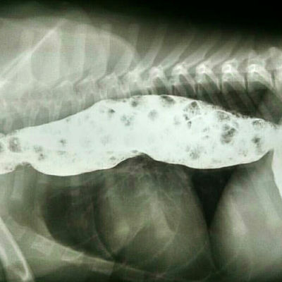
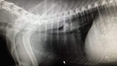
The picture above is an example of what megaoesophagus looks like on an x-ray.
That big white sausage is a dilated oesophagus with food in it.
In animals with a normal oesophagus, you cannot see it on x-ray unless there is air present in the lumen.
Sadly, due to the highly unregulated pet food industry, some pet foods have been implicated in contributing to the development of megaoesophagus.
Studies are ongoing to determine an exact cause – but we recommend that you choose pet food diets that are from credible manufacturers.
What Causes Megaoesophagus?
There are three main causes of megaoesophagus:
- Congenital (inherited) idiopathic
- Acquired idiopathic (cause unknown)
- Acquired secondary (cause known)
Congenital Acquired Megaoesophagus
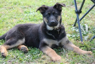
German Shepherd breeds can have an inherited form of megaoesophagus. Photo credit: Bec Mussett Photography
An increase in the incidence and heritability of megaoesophagus has been reported in the following breeds:
- Irish setter,
- Great Dane,
- German Shepherd,
- Labrador Retriever,
- Chinese Shar-Pei,
- Newfoundland,
- Wirehaired Fox Terrier
- Miniature Schnauzer
- Dachshund
Signs of congenital megaoesophagus begin to show at around 5 weeks old when pups start to be fed solid food.
These pups are often malnourished and fail to grow and thrive. Some get persistent respiratory infections such as aspiration pneumonia.
Many of these pups fail to survive, but those that do require lifelong special management.
Acquired Idiopathic Megaoesophagus
Acquired idiopathic megaoesophagus is a very frustrating disease.
It seems to occur spontaneously in dogs between 7 to 15 years old with no evidence of a cause.
Acquired Secondary Megaoesophagus
Acquired secondary megaoesophagus occurs in conjunction with another disease.
Over 25% of this type of megaoesophagus is a symptom of Myasthenia Gravis.
Other conditions that can cause megaoesophagus include:
- myopathies,
- infectious
- non-infectious
- polymyositis
- systemic lupus erythematosus
- dermatomyositis
- peripheral neuropathies,
- polyradiculoneuritis
- bilateral vagal nerve injury
- dysautonomia
- toxicity – lead, thallium, carbimide
- disorders of the neuromuscular junction,
- botulism
- tetanus
- anticholinesterase toxicity eg. organophosphate
- obstructive diseases
- strictures
- foreign bodies
- granulomas
- tumours (Spirocerca lupi)
- oesophagitis
- Central nervous system disease
- brain stem lesions
- neoplasia
- trauma
Clinical Signs Of Megaoesophagus
Animals with megaoesophagus find it difficult to hold down food.
Many dogs or cats will eat a meal and regurgitate within minutes to hours of eating.
The food may never enter the stomach and often is still in the ‘sausage shape’ of the oesophagus. This can result in these animals struggling to keep weight and look malnourished.
Frequent regurgitation can also mean these pets can suffer from pneumonia due to food entering the windpipe when they regurge.
Many animals will have a chronic cough, caused by a lung infection or irritation of bits of food in the pharynx.
You may notice that your pet has a ‘snotty nose’ or rhinitis due to food entering the nasal passages when it is regurgitated…not nice!
How Is Megaoesophagus Diagnosed?
The initial diagnosis of megaoesophagus is not hard to make via an extensive history and radiographs of the cervical and thoracic regions of your pet.
Megaoesophagus is easy to see on both plain and contrast radiographs (like the radiograph above, barium has made the oesophagus look white).
The difficulty of forming a diagnosis is determining what caused megaoesophagus in the first place.
In some circumstances, it can be a very involved process to work out why your pet has megaoesophagus.
Diagnosis of the cause of megaoesophagus can involve:
- full blood testing (including titre testing),
- further advanced imaging (fluoroscopy),
- neurological exams,
- oesophagoscopy and
- muscle biopsy and/or EMG testing.
Treatment And Management of Megaoesophagus In Dogs & Cats
It is important to treat the underlying cause of megaoesophagus in addition to supportive care.
The goals of therapy include minimising regurgitation, providing ample nutrition, and resolving and/or preventing aspiration pneumonia.
In summary:
- Resolve any primary conditions
- Medical management of other issues i.e. antibiotics for pneumonia, antacids for acid reflux
- Activity modification:
- Animals need to be fed in an upright position and kept this way for approximately 30min post eating. This utilises gravity to help food enter the stomach! Raise food height and keep the dog vertical after eating i.e. use a Bailey chair
- Nutrition – choose the right megaoesophagus dog food. This is food that is
- high calorie (these dogs tend to eat less making maintenance of weight important)
- use trial and error to test the best form of food that your dog can swallow and keep down i.e. feed a slurry vs small meatballs.
- feed small frequent meals providing time for the food to move down the oesophagus and into the stomach.
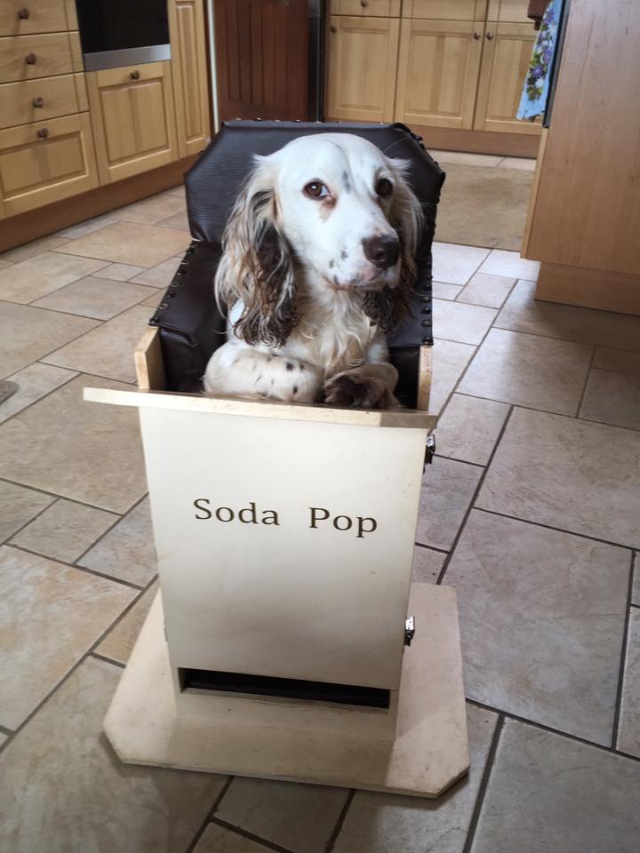
Soda Pop in her homemade Bailey Chair. Propping your dog or cat upright uses gravity to help food pass from the mouth to the stomach.
What Is The Prognosis & Life Expectancy For Dogs With Megaoesophagus?
Although megaoesophagus can be managed, it does require dedication, especially since these animals often suffer from malnutrition and aspiration pneumonia.
Some animals, if diagnosed and treated young, may grow out of the problem, however, in the majority of cases prognosis for recovery is poor.
Dedication is required to manage properly.
Do you have a pet that has megaoesophagus? Tell us how you manage their eating below in the comments section.
[easy-social-share]



Leave A Comment